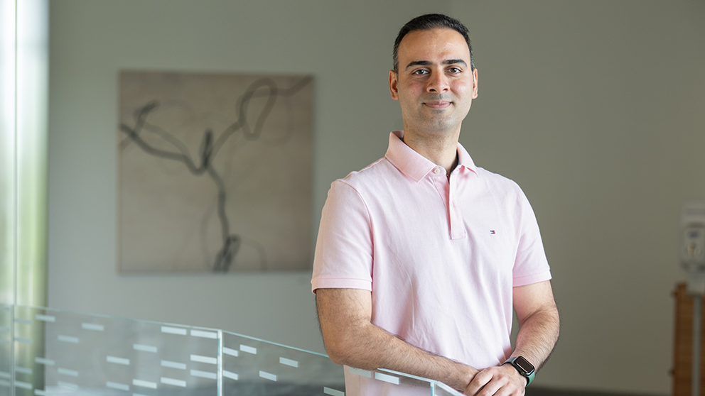
 The Jackson Laboratory's Sasan Jalili. Photo credit: Cloe Poisson
Assistant Professor Sasan Jalili, Ph.D., unites the worlds of biomedical engineering, immunology, and microbiology, offering fresh insights into the intricate interactions between the human immune system and microbiome.
Searching for synergy
A biomedical engineer by training, Jalili exhibited a passion for engineering and its potential to revolutionize healthcare from an early age. Drawn to the idea of designing tools used in hospitals, Jalili has always wanted to have a positive impact on patient outcomes. Jalili’s academic path began with a focus on creating biomaterials, natural or synthetic manufactured tools used to support, enhance or replace damaged tissue or a biological function.
Followed by a deep dive into tissue engineering and modeling human diseases across various organs, as well as engaging in therapeutic discovery studies during his Ph.D., Jalili later pursued immune engineering (a rather new discipline centered around creating tools to investigate, alter or optimize the immune system) during his postdoctoral studies. These combined experiences opened Jalili’s eyes to how many important immune cell interactions are occurring daily throughout the entire body. He is now taking a closer look at these exchanges and the symbiotic relationship between the human microbiome and the immune system.
“In the past, many people in the field of biomedical engineering have neglected the significance of immune system, especially its crosstalk with microbiome,” says Jalili. “I realized for my research to be successful I needed to have a deep understanding of immunological mechanisms of action and how we can eventually manipulate or engineer the immune system to our benefit.”
The smart band-aid
Jalili has devised two revolutionary biomedical devices, one being a microneedle skin patch. Resembling a smart band-aid, it can be applied to the skin as a non-invasive tool to longitudinally collect information about immune cells and biomarkers. Tiny projections on the surface allow the patches to be inserted on the skin and absorb biological fluid without losing their structure or perturbing the sampling area.
“These are game changing for understanding vaccine responses, autoimmunity, cancer or different systemic diseases and conditions where many important immune cell populations, such as resident memory T cells, reside in peripheral tissues like skin,” says Jalili. “These cell populations and biomarkers are often not accessed by traditional blood draws used to monitor the immune system. We are now working on ways to collect these biomarkers using our patches for the first time remotely, allowing patients to be observed without going into the clinic.”
In healthy states, the skin microbiome and innate and adaptive immune systems exist in a state of equilibrium. Jalili wants to understand how this relationship remains balanced and what causes imbalances in disease states. He hopes to discover more about the host-immune interface by sampling both microbiome and immune cells at different stages of disease.
Opening a new dimension in disease monitoring and diagnosis, the Jalili lab has obtained IRB approvals to use these patches that are made of biodegradable, FDA-approved polymer on human patients. Jalili and his team will initially be studying patient populations with autoimmune dermatological concerns, including those with psoriasis, vitiligo and dermatitis. Current methods for diagnosing these conditions often involve invasive methods such as skin biopsies. But Jalili’s microneedle patches could allow dermatologists to cut back on these drastic measures while also painting a more accurate picture of what is really happening on the skin surface and below.
“Few have approached these disease states from an engineering aspect. Now that we understand immune response, we can investigate how the skin microbiome affects this response in these dermatological diseases and vice versa,” says Jalili.
Chipping in
Another groundbreaking aspect of Jalili’s work is his development of an organ-on-chip system. The devices go far beyond the traditional cell culture methods using Petri dishes or flasks, building on patient-derived organoids or iPSCs with unprecedented capability to emulate the human cellular microenvironment and tissue function. These organ chips will allow researchers to study cells as if they were still within the body as they respond to the cues from other cells, tissue-tissue contact, as well as mechanical pressures such as vascular and lymphatic flow.
These tiny organs-on-chips replicate the complexity of human organs in a powerful yet portable package. About the size of your thumb, two channels run along the top and bottom. Cells from various tissues are cultured on both the upper and lower channels, separated with a central membrane coated with extracellular matrix (ECM). While keeping the two layers of cells separate, the membrane still allows the different cell types to communicate and interact. This arrangement replicates a tissue-tissue interface, allowing for the migration of cells. The flexible membrane and the attached tissues undergo rhythmic deformations, effectively mimicking mechanical movements at the organ level, such as breathing or peristalsis motions. The Jalili lab plans to use this organ-on-chip method to study cells in a more physiologically relevant environment, enhancing explorations of host immunity and the microbiome in the contexts of infectious disease, cancer and aging.
“If you want to study a particular cancer, we can take the cells with the specific genetic abnormality in that tumor and grow it on the chip,” says Jalili. “Then we can introduce immune cells, microbiome, and metabolites found in that organ that are essential in regulating that cancer or other associated abnormalities. We are also interested to see how changes in disease states and immunity affect the microbiome and cause dysbiosis. This tool will be pertinent for our gastrointestinal work since imbalances of gut microflora have been linked to several conditions related to the stomach and intestines such as Inflammatory bowel disease (IBD).”
Jalili’s goal is to use these devices to perform in-depth studies on host-microbiome interactions, immune responses and disease progression for gastrointestinal and skin diseases, and to expand upon the cellular modeling service JAX already offers as a research resource. He hopes to use the chip-based models to provide a more holistic understanding of infectious diseases, autoimmunity and cancer.
“I am excited to be part of JAX’s initiative to expand cellular engineering and modeling capabilities, and the fact that I have the support to do so as a junior faculty member is very motivating. I am looking forward to building a team of collaborative individuals to help bring us into this next chapter,” says Jalili.